Blogs for Patients
What You Need to Know About Advanced Medical Imaging That Guides Better Care
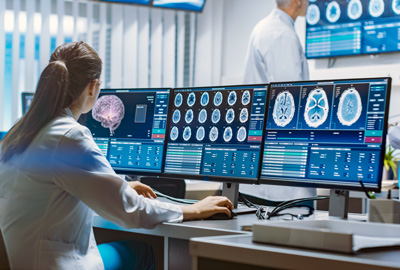
At our KC imaging center, we provide cutting-edge diagnostic tools that help physicians make accurate, timely decisions about your health. From Gastric Emptying Studies, which assess digestive function, to PET/CT scans, which play a crucial role in detecting and monitoring disease, and advanced brain imaging for diagnosing and treating Traumatic Brain Injuries (TBI), our expertise ensures you get the answers you need. Learn how these imaging technologies work, when they’re recommended, and how they support better patient outcomes.
Understanding Prostate MRI
A Patient's Guide to Advanced Cancer Detection
Prostate cancer remains the second most common malignancy in men worldwide, making early and accurate detection crucial for optimal patient outcomes. If you're facing possible prostate cancer screening or diagnosis, you've likely heard about prostate MRI. This advanced imaging test is revolutionizing how doctors detect prostate cancer, making diagnosis more accurate while helping many men avoid unnecessary procedures.
What is Prostate MRI?
Prostate MRI is a detailed imaging scan that creates precise pictures of your prostate gland. Unlike traditional methods that relied on essentially random tissue sampling, MRI allows doctors to actually see suspicious areas before deciding whether a biopsy is needed.
How Does Prostate MRI Work?
The MRI scan combines three different types of imaging to create a comprehensive picture of your prostate:
Anatomical Images: Show the detailed structure of your prostate, including its different zones and any abnormal areas.
Diffusion Images: Measure how water moves through prostate tissue. Cancer cells tend to be packed more tightly, restricting water movement in ways that show up on these images.
Contrast Images: After receiving an IV contrast injection, these images track blood flow patterns. Cancerous areas often have different blood flow compared to healthy tissue.
The entire scan typically takes 30-45 minutes and doesn't involve any radiation.
Understanding Gastric Emptying Studies
A Window Into Your Digestive Health

Gastric emptying nuclear medicine studies provide crucial insights into how efficiently your stomach empties its contents. This diagnostic exam helps identify conditions like gastroparesis, where the stomach's natural emptying process is delayed. A gastric emptying study can also determine if the stomach empties too quickly, a condition known as rapid gastric emptying.
What to Expect During Your Study
The procedure begins with eating a light meal containing a small amount of radioactive material. This special meal typically consists of eggs labeled with Technetium-99m, a safe radioactive isotope. After the meal, a one-minute image will be captured every hour for the next four hours.
Why Your Doctor May Order This Test
Common symptoms that warrant a gastric emptying study include:
- Persistent nausea and vomiting
- Early satiety (feeling full quickly)
- Upper abdominal pain
- Unexplained weight loss
- Bloating after meals
Unveiling the Power of PET/CT Imaging
Element Medical Imaging is excited to announce the addition of PET/CT imaging. PET/CT (PET Scan) is a cutting-edge diagnostic tool that combines two powerful diagnostic imaging modalities: Positron Emission Tomography (PET) and Computed Tomography (CT). This advanced technology offers an unparalleled view inside the human body, revolutionizing the way we detect and monitor various medical conditions.
Understanding PET/CT Imaging
PET/CT is a non-invasive exam that fuses the strengths of PET and CT scans. The PET portion of the exam detects areas of high metabolic activity, such as tumors or inflammation, while the CT portion provides detailed anatomical images of the body’s internal structures. By simultaneously combining the two modalities, PET/CT offers a comprehensive picture of both the structure and function of the body yielding more accurate diagnoses and personalized treatment plans.
Diagnostic Capabilities of PET/CT
PET/CT imaging plays a pivotal role in the detection, staging and monitoring of various cancers, including prostate, lung, breast, colorectal and lymphoma. Additionally, it aids in the diagnosis of neurological disorders such as Alzheimer’s and Parkinson’s disease.
The Role of Diagnostic Imaging in Traumatic Brain Injuries
Traumatic brain injury (TBI) is a significant public health concern worldwide, affecting millions of individuals each year. Whether resulting from sports-related accidents, vehicular collisions, falls or other incidents, TBIs can have profound and often life-altering consequences. Prompt and accurate diagnosis is crucial for ensuring appropriate treatment and minimizing long-term complications. In this blog post, we delve into the pivotal role of diagnostic imaging in diagnosing and treating traumatic brain injuries.
The Importance of Diagnostic Medical Imaging
 Diagnostic imaging techniques play a pivotal role in the evaluation and management of traumatic brain injuries. While clinical assessment and patient health history are essential components of diagnosis, imaging modalities provide invaluable insights into the extent and nature of brain injury. By visualizing internal structures of the brain, these techniques enable healthcare professionals to make informed decisions regarding patient care.
Types of Diagnostic Imaging for TBI
Several imaging modalities are commonly employed in the diagnosis of traumatic brain injury. These include:
- Computed Tomography (CT) Scan: CT scans are often the initial imaging modality of choice for patients with suspected TBI. CT scans provide detailed cross-sectional images of the brain, allowing healthcare providers to assess for intracranial hemorrhage, skull fractures and other acute injuries. The speed and accessibility of CT imaging make it particularly valuable in emergency settings for rapid triage and decision-making.